


Arsenic Effects in Infants and Children: A Comprehensive Review of Developmental Toxicity, Mismetallation, Microbiome Disruption,and Microbial Metallomics
Toxicity Research
Arsenic Effects in Infants and Children: A Comprehensive Review of Developmental Toxicity, Mismetallation, Microbiome Disruption,and Microbial Metallomics

Abstract
This review synthesizes evidence that arsenic exposure during fetal life and early childhood produces measurable and persistent impacts on neurodevelopment, immune function, and long-term health risk. Early-life exposure is driven predominantly by contaminated drinking water, dietary intake (notably rice and rice-based infant foods), and maternal–fetal transfer, with additional contribution via lactation depending on maternal exposure context. Because developmental systems are rapidly maturing, arsenic exposure during these windows can disrupt brain growth, cognition, and immune programming, with effects modified by sex, mixture co-exposures, and how arsenic is measured and speciated. Mechanistically, arsenic acts not only through oxidative stress and inflammation but also via mismetallation, notably displacement of zinc in zinc finger proteins, leading to protein dysfunction, proteotoxic stress, and epigenetic reprogramming. Emerging microbiome evidence indicates arsenic can reshape microbial community composition and function, reducing protective metabolites (e.g., short-chain fatty acids) and promoting barrier dysfunction and immune dysregulation. Prevention priorities center on reducing arsenic in water and infant foods, diversifying infant grains away from high-arsenic sources, strengthening surveillance and standards, and integrating microbiome-informed strategies into risk communication.
Keywords
Arsenic exposure; infants; children; prenatal transfer; developmental toxicity; cognition; mismetallation; zinc finger proteins; microbiome dysbiosis; immune dysfunction; microbial metallomics; mitigation.

Introduction
Arsenic contamination is described as a major global health threat, with over 200 million people exposed to elevated arsenic concentrations, primarily through contaminated drinking water [1]. Infants and young children are especially vulnerable because exposure can occur during critical developmental windows, and maternal-fetal transfer occurs readily across the placenta [2]. The review describes substantial placental arsenic accumulation in exposed populations compared to non-exposed individuals, underscoring the importance of prenatal exposure control [2].
Arsenic Exposure Pathways and Early-Life Vulnerability
Sources and Routes of Prenatal and Early Childhood Exposure
Arsenic contamination represents one of the most significant global public health threats, with over 200 million people exposed to elevated arsenic concentrations, primarily through contaminated drinking water [1]. Infants and young children face particular vulnerability due to their early developmental windows and enhanced toxicological sensitivity. Maternal-fetal arsenic transfer occurs readily across the placenta, with cord blood arsenic concentrations reaching levels nearly as high as maternal exposure [2]. The median placental arsenic concentration in exposed populations can reach 34μg/kg compared 7μg/kg to in non-exposed individuals, indicating substantial placental accumulation [2]. Geographic disparities in exposure are pronounced, with infants in Bangladesh, Pakistan, and India exposed to dietary arsenic levels significantly exceeding health reference values of 0.3-8μg/kg/day [1]. Early childhood exposure through breast milk presents an additional route of transmission, with breast milk arsenic levels varying considerably based on maternal dietary intake and environmental exposure [3]. The metabolic handling of arsenic differs markedly in early development compared to adults, with methylation processes showing enhanced activity during pregnancy and early infancy, converting inorganic arsenic to less immediately toxic dimethylarsinic acid (DMA) [2].
Critical Developmental Windows
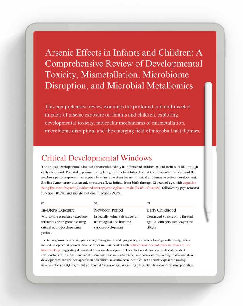
The critical developmental windows for arsenic toxicity in infants and children extend from fetal life through early childhood. Prenatal exposure during late gestation facilitates efficient transplacental transfer, and the newborn period represents an especially vulnerable stage for neurological and immune system development [4]. Studies demonstrate that arsenic exposure affects infants from birth through 12 years of age, with cognition being the most frequently evaluated neuropsychological domain (94.8% of studies), followed by psychomotor function (40.3%) and social-emotional function (29.9%) [4]. In this framework, three sensitive windows are emphasized: in-utero exposure, where mid-to-late pregnancy exposure influences brain growth during critical neurodevelopmental periods; the newborn period, which is especially vulnerable for neurological and immune system development; and early childhood, where vulnerability can continue through age 12 with persistent cognitive effects [4]. In-utero exposure to arsenic, particularly during mid-to-late pregnancy, influences brain growth during critical neurodevelopmental periods. Arsenic exposure is associated with reduced head circumference in infants at 1–3 months of age, suggesting diminished brain size development [5]. The effect size demonstrates dose-dependent relationships, with a one standard deviation increase in in-utero arsenic exposure corresponding to decrements in developmental indices [6]. Sex-specific vulnerabilities have also been identified, with arsenic exposure showing adverse effects on IQ in girls but not boys at 5 years of age, suggesting differential developmental susceptibilities [7].
Cognitive Impairment and Neuropsychological Deficits
The most extensively documented consequence of childhood arsenic exposure is cognitive impairment. A systematic review analyzing 24 studies demonstrated a consistent inverse relationship between arsenic exposure and cognitive performance in children, with higher arsenic levels associated with lower IQ scores, slower processing speeds, and impaired memory and language skills [8]. These cognitive deficits were evident across diverse geographical regions and persisted even after adjusting for sociodemographic factors. A meta-analysis of 17 observational studies involving 6,907 participants found that a 50% increase in prenatal arsenic exposure was associated with reductions of 0.51 points in the Mental Development Index (MDI) and 0.15 points in the Psychomotor Development Index (PDI), though these effects did not achieve statistical significance across all populations [6]. This apparent paradox may reflect heterogeneity in exposure measurement methods, with studies measuring total urinary arsenic without species differentiation potentially underestimating inorganic arsenic toxicity.
Neurodevelopmental Mechanisms and Molecular Pathways
Developmental arsenic exposure triggers molecular pathways that fundamentally alter brain maturation. In a maternal arsenic exposure model in mice, time-series RNA sequencing revealed significant temporal correlation between arsenic neurodevelopmental toxicity and altered hippocampal mRNA expression profiles during critical postnatal developmental windows [9]. Downregulation of genes associated with neurogenesis and telomere maintenance was observed, with critically shortened telomeres inhibiting neural stem cell proliferation, impairing neuroblast maturation, and reducing neurosphere numbers and sizes. In this context, the review emphasizes coordinated effects that include downregulation of neurogenesis and telomere maintenance genes, structural changes such as impaired dendritic complexity and altered dendritic spine morphology, and synaptic disruption with altered synaptic ultrastructure linked to cognitive impairment [9]. Arsenic exposure impaired dendritic complexity in the hippocampus, altered dendritic spine morphology, and disrupted synaptic ultrastructure, ultimately leading to cognitive impairment [9]. These structural alterations in neural tissue represent permanent modifications of brain architecture during critical windows of vulnerability. The neural stem cell population exhibited decreased stemness markers, with BrdU+ and MAP2+ cells reduced while GFAP+ cells were increased, indicating a fundamental shift in neural progenitor cell behavior that would compromise long-term cognitive capacity [9].
Multiplex Metal Exposure Effects and Synergism
Most environmental exposures involve multiple heavy metals simultaneously, with arsenic co-occurring with lead, mercury, cadmium, and other metalloids. In an artisanal gold mining region of Tanzania, prenatal multi-chemical exposure to lead, mercury, cadmium, and arsenic showed synergistic effects on developmental outcomes [10]. Joint metal exposure decreased gross motor skills by 17.78%, language ability by 55.36%, and general developmental milestones by 13.36%, with the combined effect greater than would be expected from additive exposure models [10]. Lead amplified the developmental toxicity of cadmium and arsenic, suggesting that co-occurring toxic metals potentiate their individual developmental effects beyond simple additive toxicity [10].
Arsenic-Induced Zinc Finger Protein Mismetallation
A central molecular mechanism of arsenic toxicity involves displacement of zinc ions from zinc finger proteins and other metalloproteins, causing protein misfolding, loss of function, and cellular dysfunction. Arsenite (As3+) selectively binds to cysteine-rich zinc finger motifs with C3H1 and C4 configurations, displacing the coordinated zinc ion [11]. This binding is not a simple replacement but rather induces conformational changes in the zinc finger domain structure. Arsenite exposure in human keratinocytes impaired the splicing function of ZRANB2, an alternative splicing regulator protein containing two C4 zinc finger motifs essential to its structure [11]. Within 3–24 hours of arsenic exposure, ZRANB2-dependent splicing of target mRNA was impaired, demonstrating functional consequences of zinc displacement [11]. The mismetallation mechanism extends beyond simple zinc loss. Chemoproteomic approaches using biotin-As(III) probes identified multiple nuclear arsenite-binding proteins involved in mRNA splicing, DNA repair, and replication [12]. As3+ binding to splicing factor 1 (SF1) perturbed mRNA splicing in human cells, indicating that mismetallation of splicing machinery disrupts this essential RNA processing function [12]. The selectivity of arsenite for C3H1 and C4 zinc fingers versus C2H2 zinc fingers suggests that not all zinc proteins are equally affected; rather, arsenite targets a specific subset of zinc finger configurations based on cysteine spacing and coordination geometry.
Arsenic-Induced Protein Aggregation and Proteotoxic Stress
Beyond direct zinc displacement, arsenic exposure induces protein aggregation through mismetallation-dependent mechanisms. In bacterial systems, soft metalloids including arsenic promote protein aggregation both during translation and post-translationally, disrupting multiple essential biological processes [13]. The aggregated proteins are involved in amino acid biosynthesis, energy metabolism, and other critical functions, with accumulation of aggregates directly linked to cell death. Protein Aggregation Arsenic coordinates with cysteine residues, causing proteins to misfold and aggregate Proteotoxic Stress Upregulation of chaperones and proteasomal degradation creates metabolic burden Cell Death Overwhelmed proteostatic capacity leads to cellular dysfunction and death In human systems, arsenic’s interaction with cysteine and histidine-containing proteins initiates aggregation cascades. The aggregation occurs preferentially in proteins with multiple cysteine residues that are susceptible to arsenic coordination. This proteotoxic stress activates cellular quality control mechanisms, including upregulation of molecular chaperones and proteasomal degradation pathways, representing a metabolic burden on developing tissues already stressed by arsenic exposure [14]. In cells lacking adequate chaperone systems, arsenic-induced protein aggregation overwhelms proteostatic capacity, leading to cell death.
Epigenetic Modifications and Gene Regulation Through Mismetallation
Arsenic exposure modulates gene expression through epigenetic mechanisms involving metal-responsive transcription factors. The metal-activated transcription factor 1 (MTF1) mediates induction of metallothionein genes in response to arsenic binding [15]. Arsenic binds to a C-terminal cysteine cluster of MTF1, activating its transcriptional activity and upregulating genes involved in metal homeostasis and detoxification [15]. However, this compensatory response has a metabolic cost, diverting cellular resources from normal developmental processes [15]. Arsenic exposure also reduces global histone H4 acetylation at lysine 16 through direct binding to histone acetyltransferase hMOF, inhibiting its catalytic activity [16]. This epigenetic modification alters chromatin structure and gene expression patterns across multiple genes, potentially affecting genes essential for neurodevelopment and immune function [16]. The altered methylation patterns persist beyond the exposure period, suggesting long-term epigenetic dysregulation from developmental arsenic exposure [17].
Microbiome Composition Shifts in Response to Arsenic Exposure
Arsenic exposure profoundly alters microbial community composition in infant and child gut microbiota. Prenatal arsenic exposure in murine models altered the fecal microbiome composition, with significant decreases in Firmicutes abundance in arsenic-exposed offspring [18]. Functional analysis revealed that arsenic exposure shifted genes involved in crucial metabolic pathways such as insulin signaling and non-alcoholic fatty liver disease pathways, suggesting that arsenic-induced microbiome dysbiosis could predispose to metabolic disease [18]. In human populations exposed to arsenic through contaminated drinking water, the microbiota exhibits characteristic dysbiotic shifts. Food chain microbiome studies examining responses to arsenic across multiple ecosystem components revealed that chemical stressors, including arsenic, decreased microbiome diversity in soil but caused compositionally distinctive shifts in water, sediment, plant, and animal microbiomes [19]. The dysbiotic communities became compositionally more similar to each other in response to stress, suggesting convergence toward a stress-selected community composition [19]. Importantly, different bacterial taxa responded specifically to arsenic exposure, with stochastic effects particularly notable in host-associated communities [19].
Functional Consequences of Microbiome Dysbiosis
Beyond compositional changes, arsenic-induced microbiota dysbiosis impairs critical bacterial metabolic functions. In arsenic-contaminated groundwater systems, distinct microbial communities enriched in response to varying electron acceptor regimes included arsenite-oxidizing bacteria (Deinococcus), nitrate-reducing bacteria (Denitratisoma), sulfate-reducing bacteria (Macellibacteroides), and other functional groups [20]. However, the metabolic plasticity of these communities was constrained by arsenic stress, leading to functional narrowing. Heavy metal exposure alters functional genes related to xenobiotic metabolism and amino acid biosynthesis, reducing the microbiota’s capacity to synthesize essential metabolites [21]. Dysbiotic microbiota also show reduced abundance of genes involved in short-chain fatty acid production, potentially compromising intestinal barrier integrity and immune development in infants. The dysbiotic microbiota shows impaired capacity for critical metabolic functions, and this functional impairment extends beyond simple reduction in community diversity, with specific metabolic pathways preferentially affected.
Microbiota-Mediated Barrier Dysfunction and Immune Dysregulation
Arsenic-induced microbiota dysbiosis contributes to intestinal barrier dysfunction and systemic immune dysregulation. In a preclinical study, arsenic-induced oxidative stress and barrier dysfunction in gut epithelial cells was mitigated by the microbial metabolite urolithin A (UroA), a polyphenol metabolite produced by beneficial bacteria [22]. This finding demonstrates that dysbiosis-related loss of metabolite-producing bacteria exacerbates arsenic toxicity. The dysbiotic microbiota lacks bacteria capable of producing protective metabolites, leaving the epithelium vulnerable to arsenic-induced apoptosis and tight junction protein disruption. The dysbiotic microbiota exhibits altered production of metabolites that regulate immune development. Short-chain fatty acids produced by fermentative bacteria modulate immune tolerance and promote anti-inflammatory responses, processes critical for proper immune development in infants [23]. Arsenic-induced dysbiosis reduces butyrate-producing bacterial populations, compromising the production of these essential signaling molecules. This metabolic dysfunction contributes to immune dysregulation, potentially explaining the increased susceptibility to infections observed in arsenic-exposed children.
Microbial Arsenic Metabolism Pathways and Gene Expression
Microorganisms inhabiting arsenic-contaminated environments express genes encoding arsenic metabolism enzymes that influence arsenic bioavailability and toxicity. Arsenite-oxidizing bacteria oxidize toxic As3+ to less mobile As5+ through arsenite oxidase (aioA gene products), while arsenate-reducing bacteria convert As5+ back to As3+ through arsenate reductase (arrA gene products) [24]. In arsenic-contaminated groundwater in Bangladesh, 72 isolated bacterial strains harbored diverse arsenotrophic genes: 23 isolates possessed the arsenite efflux pump gene (arsB) with high abundance, and 10 isolates harbored the arsenite oxidase gene (aioA). Proteobacteria, Firmicutes, and Acidobacteria dominated the arsenotrophic communities, with genera including Pseudomonas, Acinetobacter, Stenotrophomonas, Achromobacter, Paraburkholderia, Comamonas, and Klebsiella identified as potential arsenic detoxifiers. The metabolic capacity of the microbiota for arsenic transformation creates a dynamic system of arsenic speciation. Rhizosphere microbiota of arsenic-hyperaccumulator plants (Pteris vittata) engaged in coordinated arsenic speciation through multiple metabolic processes [25]. The rhizosphere community was dominated by Proteobacteria, Acidobacteriota, and Ascomycota, with 44 bacterial and 10 fungal genera identified as core microorganisms capable of arsenic metabolism. Microbial-mediated arsenic methylation and reduction processes, coupled with carbon fixation, sulfur oxidation, and phosphorus mineralization, contributed to an “As-multielement cycling” synergy that enhanced plant arsenic uptake [25].
Mitigation Strategies and Bioremediative Approaches
Environmental and Dietary InterventionsEvidence-based strategies for reducing arsenic exposure in infants and children include improved agricultural practices, dietary modifications, and regulatory interventions. The comprehensive review of arsenic exposure in infant and child diets identified critical sources including rice and rice-based products, infant cereals, and contaminated groundwater [1]. Geographic disparities in regulatory frameworks were highlighted, with recommendations for stricter regulatory limits on arsenic in infant products and encouragement of low-arsenic dietary alternatives. Dietary interventions targeting arsenic reduction in infants include promotion of diverse grain sources and modified feeding practices. Rice-based infant cereals present particular concern due to rice’s propensity for arsenic accumulation [29]. Heavy metal contamination in commonly used thickeners for infants with reflux or dysphagia was evaluated in 56 infants less than one year of age, with urinary arsenic concentrations assessed across different thickener types. Infants with higher servings of alternative arsenic sources via solid foods were more likely to have higher urinary arsenic levels, suggesting that cumulative dietary arsenic from multiple sources represents a significant exposure pathway.
Microbial-Based Bioremediative ApproachesMicrobial bioremediation represents a promising strategy for reducing arsenic bioavailability in contaminated environments. Arsenic-oxidizing bacteria (AOB) isolated from highly contaminated mining soils can transform toxic As3+ into less mobile As5+ [30]. Acinetobacter sp. TMKU7 isolated from arsenic-contaminated soil transformed 80% of As3+ to As5+ under culture conditions and expressed plant growth-promoting traits including siderophore production and indole-3-acetic acid synthesis. The arsenite oxidase enzyme was constitutively expressed and localized to the periplasmic fraction, with partially purified enzyme showing Km of 41.43 μM and Vmax of 0.19 μM min−1 g−1 protein [30]. Bioaugmentation with specific arsenic-resistant bacterial strains enhanced arsenic mobility and removal from contaminated soils. In highly arsenic and antimony-contaminated Slovak mining soils, biostimulation and bioaugmentation with Cupriavidus metallidurans and Cupriavidus oxaleticus resulted in mean bleached arsenic fractions of 37.6% and 41.3%, respectively [31]. Consortium-based approaches combining multiple bacterial strains with complementary metabolic capacities showed enhanced remediation efficiency compared to single-strain inoculants.
Integrated Public Health Strategies and Future Directions
Comprehensive public health approaches addressing arsenic exposure in infants and children require integration of environmental monitoring, dietary regulation, and community awareness. The FDA has received recommendations to re-evaluate permissible limits of arsenic in cereals and juices aimed at children consumption, with proposed revisions emphasizing protection of the most vulnerable populations [32]. Current FDA standards allow up to 100 μg/L of arsenic in apple juice, a level that may exceed safe exposure for young children given their higher per-kilogram intake relative to body weight [32]. Policy interventions must account for geographic disparities in arsenic exposure and vulnerability. In Bangladesh, Pakistan, and India, where dietary arsenic exposure significantly exceeds health reference values, targeted interventions are most urgently needed [1]. These include groundwater treatment infrastructure development, agricultural practices modification to reduce arsenic uptake, and fortification of foods with low-arsenic sources [1]. Community-based environmental monitoring programs provide essential data for informed decision-making regarding remediation priorities. Community-awareness programs must address maternal knowledge of arsenic exposure risks during pregnancy and lactation, given the importance of early life windows. Comprehensive information regarding arsenic in drinking water, food sources, and occupational exposures would enable informed maternal decision-making to reduce fetal and early childhood exposure. The integration of microbiome science into public health messaging, emphasizing the protective role of beneficial bacteria and the consequences of dysbiosis from environmental toxin exposure, could support development of microbiota-protective strategies including dietary diversity and reduced antimicrobial exposure.
Discussion
Arsenic exposure in early life emerges as a developmental risk primarily because exposure can begin before birth and continue through infancy and childhood, overlapping with periods of rapid brain and immune system maturation. The findings summarized in this review indicate that the strongest and most consistently reported human outcome is reduced neurodevelopmental performance, with multiple studies linking higher arsenic exposure to poorer cognitive metrics across childhood. Although effect sizes vary between cohorts, the overall pattern is directionally consistent, and differences in exposure measurement, including whether arsenic is speciated, likely contribute to heterogeneity across studies.
Mechanistic evidence in the review strengthens biological plausibility for these developmental associations. Experimental work describing altered hippocampal transcriptional profiles, impaired neurogenesis, telomere-related disruption, and lasting changes in dendritic and synaptic structure provides coherent pathways through which early arsenic exposure can translate into persistent cognitive effects. The review also emphasizes that arsenic toxicity is not limited to classic oxidative and inflammatory injury; it includes a distinctive molecular mechanism in which arsenite displaces zinc from specific zinc finger proteins, disrupting essential processes such as mRNA splicing and contributing to protein misfolding, aggregation, and proteotoxic stress. In parallel, arsenic-associated epigenetic alterations, including changes in histone acetylation and persistent methylation patterns, suggest that some exposure effects may be durably programmed beyond the exposure period.
A further implication is that arsenic exposure operates within real-world mixtures. Evidence summarized in this review indicates that co-exposure with other toxic metals can produce synergistic developmental harms, with lead described as amplifying cadmium and arsenic toxicity, reinforcing the need for risk management approaches that address combined exposures rather than single metals in isolation. The microbiome component adds another layer of complexity: arsenic-associated dysbiosis and functional shifts, including reduced microbial capacity for key metabolic outputs such as short-chain fatty acids, may weaken barrier integrity and contribute to immune dysregulation, potentially increasing vulnerability to infection and other downstream outcomes in exposed children.
From a public health perspective, the review supports a prevention-first posture because key exposure sources are modifiable. Evidence-based interventions combining environmental remediation through arsenic-resistant microorganisms, dietary modifications reducing arsenic exposure, regulatory limits on arsenic in infant foods, and community-based awareness programs offer practical pathways to reduce developmental burden. Future research priorities described in this review, including longitudinal studies with microbiota profiling, mechanistic studies of dysbiosis and mismetallation, and intervention trials of microbiota-protective strategies, could improve causal understanding and inform more targeted prevention approaches for vulnerable populations.
References
S. L et al., “A comprehensive review on arsenic exposure and risk assessment in infants and young children diets: Health implications and mitigation interventions in a global perspective.” 2025.
M. Vahter, “Health effects of early life exposure to arsenic,” Wiley, 2008.
G. Concha, G. Vogler, D. Lezcano, B. Nermell, and M. Vahter, “Exposure to inorganic arsenic metabolites during early human development,” Oxford University Press, 1998.
A. Kumar et al., “High arsenic contamination in the breast milk of mothers inhabiting the gangetic plains of bihar: A major health risk to infants,” Environmental Health, 2024.
A. Signes-Pastor et al., “Infants dietary arsenic exposure during transition to solid food,” Scientific Reports, 2018.
T. Y, H. Q, Z. M, G. E, and W. Y, “Exposure to arsenic and cognitive impairment in children: A systematic review.” 2025.
S. R, J. Y, L. W, D. W, and F. L, “Effects of prenatal arsenic, cadmium, and manganese exposure on neurodevelopment in children: A systematic review and meta-analysis.” 2025.
N.-B. L, C.-G. LM, B. JA, V. J, K. MR, and S.-P. AJ, “Arsenic exposure and neuropsychological outcomes in children: A scoping review.” 2025.
L. SL et al., “Early postnatal and concurrent exposure to metals and neurobehavioral outcomes at 5 years: Associations with individual environmental exposures and mixtures.” 2024.
T. C et al., “Dynamic telomere length response to neurodevelopmental arsenic exposure: Insights into transcriptional regulation and neuronal morphogenesis.” 2025.
C. R. Tyler, J. J. W. Smoake, E. R. Solomon, E. Villicana, K. Caldwell, and A. Allan, “Sex-dependent effects of the histone deacetylase inhibitor, sodium valproate, on reversal learning after developmental arsenic exposure,” Frontiers in Genetics, 2018.
S. Htway, T. Suzuki, S. Kyaw, K. Nohara, and T.-T. Win-Shwe, “Effects of maternal exposure to arsenic on social behavior and related gene expression in F2 male mice,” Environmental Health and Preventive Medicine, 2021.
R. Soler-Blasco et al., “Genetic susceptibility to neurotoxicity related to prenatal inorganic arsenic exposure in young spanish children.” Environmental Science and Technology, 2023.
L. Beaver et al., “Combinatorial effects of zinc deficiency and arsenic exposure on zebrafish (danio rerio) development,” PLoS ONE, 2017.
S. Himeno, D. Sumi, and H. Fujishiro, “Toxicometallomics of cadmium, manganese and arsenic with special reference to the roles of metal transporters,” Toxicological Research, 2019.
E. F. Winterbottom et al., “The aquaglyceroporin AQP9 contributes to the sex-specific effects of in utero arsenic exposure on placental gene expression,” Environmental Health, 2017.
H. Zhang et al., “Association and mediation analyses among multiple metal exposure, mineralocorticoid levels, and serum ion balance in residents of northwest china,” Scientific Reports, 2024.
H. YH et al., “Combined effects of global DNA methylation, blood lead and total urinary arsenic levels on developmental delay in preschool children.” 2025.
G. Singh, S. O. Aftab, and O. Dhankher, “Arabidopsis thaliana oxoprolinase 1 (AtOXP1) maintains glutamate homeostasis, promotes arsenite and mercury tolerance, and reduces accumulation in plants.” The Plant Journal, 2025.
A. J. A.-A. Oudah, R. W. S. AL-Jebory, A. H. R. Al.Zurfi, and H. M. Jasim, “Mechanisms of heavy metal toxicity at the cellular, molecular and general health levels,” Journal of Biomedicine and Biochemistry, 2025.
J. M. Miranda et al., “Analysis of gut bacteriome of in utero arsenic-exposed mice using 16S rRNA-based metagenomic approach,” Frontiers in Microbiology, 2023.
K. Gokulan et al., “Exposure to arsenite in CD-1 mice during juvenile and adult stages: Effects on intestinal microbiota and gut-associated immune status,” mBio, 2018.
C. Zhang et al., “Unexpected genetic and microbial diversity for arsenic cycling in deep sea cold seep sediments,” bioRxiv, 2023.
A. AriasBorrego, M. SelmaRoyo, M. C. Collado, N. Abril, and T. GarcaBarrera, “Impact of chemical cocktails exposure in shaping mice gut microbiota and the role of selenium supplementation combining metallomics, metabolomics, and metataxonomics,” Elsevier BV, 2022.
S. M. Ahmed et al., “A prospective cohort study of in utero and early childhood arsenic exposure and infectious disease in 4- to 5-year-old bangladeshi children,” Environmental Epidemiology, 2020.
D. D and S. MH, “Prenatal and childhood exposures to heavy metals and their associations with child cognition, motor skills, behaviour and mental health.” 2025.
M. P. Coryell, B. A. Roggenbeck, and S. T. Walk, “The human gut microbiomes influence on arsenic toxicity,” Springer Science+Business Media, 2019.
Z.-B. Ge et al., “Two-tiered mutualism improves survival and competitiveness of cross-feeding soil bacteria,” Springer Nature, 2023.
K. Zbieralski et al., “Multilevel regulation of membrane proteins in response to metal and metalloid stress: A lesson from yeast,” International Journal of Molecular Sciences, 2024.
N. Jamunasri, A. Iyer, M. L. Prabha, R. Issac, and S. Murugan, “Metalloproteomics: Unraveling the metal binding proteins of diverse metal-resistant bacteria,” Asian Journal of Chemistry, 2024.
I. Seregin and A. Kozhevnikova, “Phytochelatins: Sulfur-containing metal(loid)-chelating ligands in plants,” International Journal of Molecular Sciences, 2023.
S. Khullar and M. S. Reddy, “Cadmium and arsenic responses in the ectomycorrhizal fungus laccaria bicolor: Glutathione metabolism and its role in metal(loid) homeostasis,” Environmental Microbiology Reports, 2018.
W. Maret, “The quintessence of metallomics: A harbinger of a different life science based on the periodic table of the bioelements,” Oxford University Press, 2022.
K. M. Wai et al., “In-utero arsenic exposure and growth of infants from birth to 6 months of age: A prospective cohort study in rural bangladesh,” International Journal of Environmental Health Research, 2020.
N. EC, M. RJ, A. M, T. DSK, and K. AP, “Effects of prenatal lead, mercury, cadmium, and arsenic exposure on children’s neurodevelopment in an artisanal small-scale gold mining area in northwestern tanzania using a multi-chemical exposure model.” 2025.
A. S. Ettinger, “Maternal arsenic exposure in relation to maternal and child adiposity and risk factors for diabetes,” Lippincott Williams & Wilkins, 2009.
D. S and R. DM, “Heavy metals in umbilical cord blood: Effects on epigenetics and child development.” 2024.
Citation
Pendergrass, K. Arsenic Effects in Infants and Children: A Comprehensive Review of Developmental Toxicity, Mismetallation, Microbiome Disruption,and Microbial Metallomics. Heavy Metal Tested and Certified. Published March 1, 2026.
Case Studies You May Be Interested In
 Cadmium Effects in Infants and Children: A Comprehensive Review of Health Impacts, Microbiome Shifts, and Microbial Metallomics
Toxicity Research
March 02, 2026
 Likelihood of Pear, Kale, and Spinach Purée Passing HMTc Standards: An Analysis
INFANT AND CHILD FOOD
February 22, 2026